Femoral Neck Ward’s Triangle Bone Mineral Density: a Forgotten Indicator of Bone Loss
Abstract
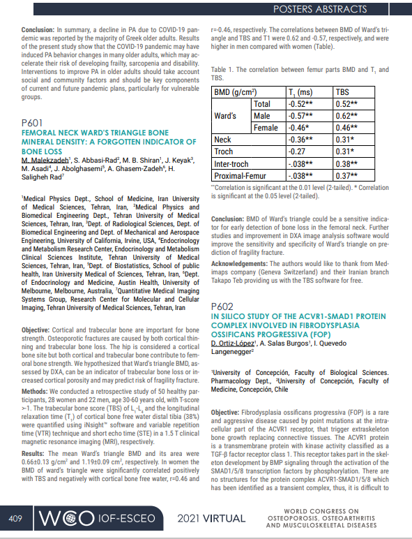
Cortical and trabecular bone both contribute to femoral strength, and osteoporotic fractures arise from cortical thinning and trabecular loss. We hypothesized that Ward’s triangle BMD—typically measured by DXA at a predominantly cortical site—may reflect trabecular deterioration or increased cortical porosity and thus predict fragility fracture risk. In a retrospective study of 50 healthy volunteers (28 women, 22 men; age 30–60 years; T-score > –1), we assessed lumbar trabecular bone score (TBS) using TBS Insight™ software and quantified cortical‐bone free‐water relaxation time (T₁) at the distal tibia (38% site) via variable‐repetition‐time/short‐echo‐time MRI on a 1.5 T clinical scanner. The mean Ward’s triangle BMD was 0.66 ± 0.13 g/cm² (area 1.19 ± 0.09 cm²). In women, Ward’s triangle BMD correlated positively with lumbar TBS (r = 0.46) and inversely with cortical‐bone free‐water T₁, indicating that lower BMD at this site reflects both trabecular loss and increased cortical porosity. These findings suggest that DXA‐derived Ward’s triangle BMD could serve as a sensitive early indicator of femoral neck bone loss. Further refinement of DXA image‐analysis software may enhance its sensitivity and specificity for predicting fragility fractures.